History
Systemic lupus erythematosus (SLE) is a chronic autoimmune disease that can affect almost any organ system. Its presentation and course are highly variable, ranging from indolent to fulminant.
A meta-analysis that reviewed the clinical manifestations of childhood-onset and adult-onset SLE found that Raynaud pleuritis and sicca were twice as common in adults as in children and adolescents. [1] In contrast, the following manifestations were statistically significantly more common in childhood-onset SLE:
-
Malar rash
-
Ulcers/mucocutaneous involvement
-
Kidney involvement, proteinuria, urinary cellular casts
-
Seizures
-
Thrombocytopenia
-
Hemolytic anemia
-
Fever
-
Lymphadenopathy
The classic presentation of a triad of fever, joint pain, and rash in a woman of childbearing age should prompt investigation into the diagnosis of SLE. [2, 3] However, patients may present with any of the following types of manifestations [4] :
-
Constitutional
-
Musculoskeletal
-
Dermatologic
-
Renal
-
Neuropsychiatric
-
Pulmonary
-
Gastrointestinal
-
Cardiac
-
Hematologic
In patients with suggestive clinical findings, a family history of autoimmune disease should raise further suspicion of SLE.
Constitutional
Fatigue, fever, arthralgia, and weight changes are the most common symptoms in new cases or recurrent active SLE flares. Fatigue, the most common constitutional symptom associated with SLE, can be due to active SLE, medications, lifestyle habits, or concomitant fibromyalgia or affective disorders.
SLE-specific fatigue or fever generally occurs in concert with other clinical markers. Fever may reflect active SLE, infection, and reactions to medications (ie, drug fever). Always exclude an infectious etiology; patients with SLE are considered immunocompromised and are therefore at higher risk for developing infections and complications. Most infections are bacterial in origin, but clinicians should always consider the possibility of atypical and opportunistic infections, particularly when these individuals are receiving immunomodulating or immunosuppressive therapy. For example, prednisone doses higher than 15 mg/day and use of methylprednisolone pulses have been associated with increased risk of severe infection. [90]
Careful history taking may help differentiate between the potential causes of fatigue or fever. Note that an acute infectious process may also trigger SLE and that the two can occur concomitantly.
Weight loss may occur in patients with active SLE. Weight gain may also be due to corticosteroid treatment or active disease, such as nephrotic syndrome (with anasarca) or myocarditis.
Musculoskeletal
Joint pain is one of the most common reasons for the initial clinical presentation of patients with SLE. Arthralgia, myalgia, and frank arthritis may involve the small joints of the hands, wrists, and knees (usually symmetrical, polyarticular). In contrast to rheumatoid arthritis, SLE arthritis or arthralgia may be asymmetrical, with pain that is disproportionate to swelling.
SLE arthropathy is rarely erosive or deforming. Characteristic hand deformities are swan neck deformities that result from recurrent synovitis and inflammation of the joint capsule, tendons, and ligaments. These deformities are usually reducible and nonerosive (resembling Jaccoud arthropathy, which is a nonerosive arthritis following acute rheumatic fever).
Another important consideration is the increased prevalence of avascular necrosis (AVN) in the SLE population relative to healthy individuals. It may be due to SLE pathogenesis and/or concomitant heavy steroid use. [91] Asymptomatic AVN is seen in up to 44% of SLE patients in the first year of therapy with high-dose corticosteroids. The most commonly affected site is the femoral head. [91] Independent risk factors for AVN in patients with SLE include the use of glucocorticosteroid or cytotoxic agents and the presence of arthritis. [92]
Dermatologic
Cutaneous manifestations of SLE include malar rash, photosensitivity, and discoid lupus. Malar rash [93] is characterized by erythema over the cheeks and nasal bridge (but sparing the nasolabial folds, which is in contrast to the rash of dermatomyositis) (see the image in Physical Examination). It lasts from days to weeks and is occasionally painful or pruritic.
Photosensitivity in SLE may be either acute or chronic. [93] The history of photosensitivity may be elicited from patients by asking if they have had any unusual rash or symptom exacerbation after sun exposure, with expected duration of approximately 2 days in classic cases.
Discoid lupus is a chronic lupus rash. [93] Discoid lesions often also develop in sun-exposed areas but are plaquelike in character, with follicular plugging and scarring. They may be part of systemic lupus or may represent discoid lupus without organ involvement, which is a separate diagnostic entity. Discoid lesions can develop in up to 25% of patients with SLE; a small case series suggested that the presence of such lesions may indicate milder disease or less kidney involvement. [94] In another review, it was reported that patients with discoid lesions rarely progressed to systemic SLE disease; there is a 5% risk of discoid lupus disease developing into the systemic condition. [95]
Subacute cutaneous lupus is a rash seen in up to 10% of SLE cases, but importantly, 50% of patients with this condition will have it in isolation without systemic lupus. [93] The characteristic lesion appearance is an annular or psoriaform patch with crusted margins. Lesions often occur on the limbs or torso in sun-exposed areas. Alopecia is an often less specific cutaneous feature of SLE. It often affects the temporal regions or creates a patchy pattern of hair loss.
Other cutaneous manifestations related to, but not specific to, SLE include the following:
-
Raynaud phenomenon
-
Livedo reticularis
-
Panniculitis (lupus profundus)
-
Bullous lesions
-
Vasculitic purpura
-
Telangiectasias
-
Urticaria
Renal
The kidney is the most commonly involved visceral organ in SLE. Although only approximately 50% of patients with SLE develop clinically evident kidney disease, biopsy studies demonstrate some degree of renal involvement in most patients. [96] Therefore, it is important to correctly classify the extent of renal involvement in SLE to improve the correlation between histologic findings and the prognosis of the kidney disease (see Biopsies and Histologic Features under Workup).Glomerular disease usually develops within the first few years of SLE onset and is often asymptomatic.
Acute kidney injury or chronic kidney disease may cause symptoms related to uremia and fluid overload. Acute nephritic disease may manifest as hypertension and hematuria. Nephrotic syndrome may cause edema, weight gain, or hyperlipidemia.
For additional information, see the Medscape article Lupus Nephritis.
Neuropsychiatric
The CNS lupus nomenclature has been revised to catalog many manifestations. [97, 98, 99] Because of the difficulty distinguishing causal SLE associations with some neurologic symptoms, only seizure and psychosis were typically included in the diagnostic criteria. Seizures related to SLE may be generalized or partial and may precipitate status epilepticus. Psychosis may manifest as paranoia or hallucinations.
However, the American College of Rheumatology (ACR) created standardized case definitions and diagnostic testing recommendations for 19 neuropsychiatric syndromes in SLE, including seizures/seizure disorders and psychosis. [100] The remainder of the neuropsychiatric syndromes are as follows [100] :
-
Acute confusional state
-
Acute inflammatory demyelinating polyradiculoneuropathy (Guillain-Barre syndrome)
-
Anxiety disorder
-
Aseptic meningitis
-
Autonomic disorder
-
Cerebrovascular disease
-
Cognitive dysfunction
-
Cranial neuropathy
-
Demyelinating syndrome
-
Headache
-
Mononeuropathy (single/multiplex)
-
Mood disorders
-
Movement disorder (chorea)
-
Myasthenia gravis
-
Myelopathy
-
Plexopathy
-
Polyneuropathy
Delirium represents a spectrum of fluctuating altered consciousness characteristic of SLE. Delirium may be caused by CNS vasculitis, encephalopathy, cerebritis, or the manifestations previously called organic brain syndrome. Aseptic meningitis, myelopathy, optic neuropathy, or other demyelinating disorders may also require urgent evaluation.
Transverse myelitis with spastic paraparesis and sensory loss at a given level is a rare but severe complication of SLE or antiphospholipid antibody syndrome. Stroke and transient ischemic attack (TIA) may be related to antiphospholipid antibody syndrome or SLE vasculitis. Posterior reversible encephalopathy syndrome (PRES) is, as the name implies, a reversible encephalopathy linked to hypertension that even may be a presenting feature for young SLE patients. [101]
Cognitive disorders may be variably apparent in many patients with SLE. Formal neuropsychiatric testing reveals deficits in 21-67% of patients with SLE. Whether this represents true encephalopathy, neurologic damage, medication effects, depression, or some other process is unclear. A 2010 multicenter study found that depression was associated with significantly poorer cognitive function in 111 patients newly diagnosed with SLE. [102]
Migraine headaches may be linked to antiphospholipid syndrome. Headache and mood disorders may be the most commonly reported neurologic manifestation of SLE, but cause and effect may be difficult to distinguish.
Acute psychiatric manifestations in CNS lupus should be considered as a diagnosis of exclusion in an SLE patient.
For additional information, see the Medscape Reference article Neurologic Manifestations of Systemic Lupus Erythematosus.
Pulmonary
Pulmonary features of SLE may manifest acutely or indolently, representing a spectrum of SLE complications. SLE may lead to multiple pulmonary complications, including pleurisy, pleural effusion, pneumonitis, pulmonary hypertension, and interstitial lung disease. The chronic steroids prescribed to patients also place them at increased risk for atypical infections.
Pleuritis is one of the formal diagnostic criteria for SLE, and it can induce chest pain and a pleural effusion. The pleural effusion in lupus is exudative, with an elevated lactate dehydrogenase level. Pleurisy with pleuritic chest pain with or without pleural effusions is the most common feature of acute pulmonary involvement in SLE. Shortness of breath or dyspnea may be due to many causes. Pulmonary embolism, lupus pneumonitis, chronic lupus interstitial lung disease, pulmonary hypertension, complement-mediated pulmonary leukoaggregation, alveolar hemorrhage, or infection may be related to lupus disease.
Most seriously, hemoptysis may herald diffuse alveolar hemorrhage, a rare, acute, life-threatening pulmonary complication of SLE.
Gastrointestinal
In general, gastrointestinal symptoms secondary to SLE are less common than adverse effects of medication or nonspecific complaints. Special consideration should be given to infectious causes (bacterial, viral [eg, CMV]), because of immunosuppression. Nausea and dyspepsia are common symptoms in patients with active SLE and are sometimes difficult to correlate with objective evidence of gastrointestinal involvement. Peptic ulcer disease is a common complication, especially in SLE patients treated with nonsteroidal anti-inflammatory agents (NSAIDs) and glucocorticoids. [103]
Occasionally, abdominal pain in active SLE may be directly related to active lupus, including peritonitis, pancreatitis, mesenteric vasculitis, and bowel infarction. Rarely, lupus enteritis may be the initial manifestation of SLE. Abdominal ultrasound can be a reliable first-line diagnostic tool in lupus enteritis, aiding early diagnosis of potentially life-threatening complications. [104] Jaundice due to autoimmune hepatobiliary disease may also occur.
Cardiac
Heart failure or chest pain must be carefully assessed in patients with SLE. Pericarditis is the most common cardiac feature of SLE, manifesting as positional chest pain that is often relieved when the patient leans forward. Myocarditis may occur in SLE with heart failure symptoms. Pulmonary hypertension may present with indolent chest pain or dyspnea.
Coronary vasculitis manifesting as angina or infarction is rarely reported. Libman-Sacks endocarditis is noninfectious but may manifest as symptoms similar to those of infective endocarditis in patients with SLE or antiphospholipid syndrome. More commonly, accelerated ischemic coronary artery disease (CAD) is associated with SLE and may present indolently as atypical anginal equivalents.
Hematologic
A history of multiple cytopenias such as leukopenia, lymphopenia, anemia, or thrombocytopenia may suggest SLE, among other etiologies, such as medication-related cytopenias. Leukopenia and, more specifically, lymphopenia are common in SLE; this, coupled with immunosuppression, may predispose persons with SLE to frequent infections.
Anemia is occasionally overlooked in young menstruating women, and a history of lymphopenia may be overlooked. Thrombocytopenia may be mild or part of a full thrombotic thrombocytopenic purpura (TTP)–like syndrome or antiphospholipid antibody syndrome. A history of recurrent early miscarriages or a single late pregnancy loss may be clues to lupus or isolated antiphospholipid antibody syndrome. [105]
Physical Examination
Almost any organ system can be involved in active SLE. The constellation of several physical findings may suggest a diagnosis of SLE. The European League Against Rheumatism/American College of Rheumatology (EULAR/ACR) diagnostic criteria are discussed in Workup. Examination findings are discussed by system. [4]
Fever is a challenging problem in SLE. It can be a manifestation of active lupus, infection, malignancy, or a drug reaction. Low-grade fever is observed in patients on immunosuppressive agents, and lymphadenopathy or splenomegaly may be found.
In patients with fever, infectious causes—both viral and bacterial—need to be ruled out. Lupus patients may be functionally asplenic and may be at risk for encapsulated bacterial infections such as meningococcemia. Patients with SLE who are on immunosuppressive therapy are at a higher risk of death due to viral infection (eg, herpes simplex virus [HSV], cytomegalovirus [CMV], varicella-zoster virus [VZV]) and should be treated accordingly if an infection is suspected. [106] An infection can mimic a lupus flare, and delays in diagnosis and treatment can increase the risk of mortality. [107]
A postdiagnostic 5-year follow-up study showed that males had a higher prevalence of thromboses, nephropathy, strokes, gastrointestinal symptoms, and antiphospholipid syndrome and that females were more likely to present with arthralgia, hair loss, Raynaud syndrome, and photosensitivity. [108] In addition, male patients were more likely to present with tendonitis, myositis, nephropathy, and respiratory tract infections.
Skin and mucous membrane findings
Malar rash is a fixed erythema that typically spares the nasolabial folds. It is a butterfly-shaped rash that can be flat or raised over the cheeks and bridge of the nose.
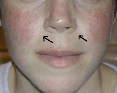 Systemic lupus erythematosus (SLE). The classic malar rash, also known as a butterfly rash, with distribution over the cheeks and nasal bridge. Note that the fixed erythema, sometimes with mild induration as seen here, characteristically spares the nasolabial folds (nasolabial fold sparing is indicated by black arrows).
Photosensitive rash is often macular or diffusely erythematous in sun-exposed areas of the face, arms, or hands and generally persists for more than 1-2 days (see the image below).
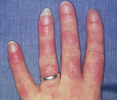 Photosensitive systemic lupus erythematosus (SLE) rashes typically occur on the face or extremities, which are sun-exposed regions. Although the interphalangeal spaces are affected, the metacarpophalangeal (MCP) and proximal interphalangeal (PIP) and distal interphalangeal (DIP) joints are spared. Photo courtesy of Dr. Erik Stratman, Marshfield Clinic.
Discoid rash occurs in 20% of patients with SLE and can result in disfiguring scars. The discoid rash can present as erythematous patches with keratotic scaling over sun-exposed areas of the skin. Follicular plugging may create scarring that may be well demonstrated in the ears. Systemic manifestations of SLE may be absent (ie, limited discoid lupus).
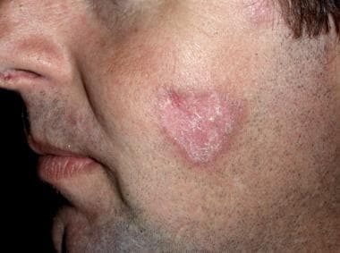 Discoid lupus erythematosus is a chronic scarring skin condition causing scaly plaques on the scalp, face, and ears.
Lupus should be considered in all patients who experience oral, or less frequently, vaginal ulcers; ulcers classically occur more than 3 times per year and are painless. Palatal ulcers are most specific for SLE.
Many other cutaneous findings are not explicitly diagnostic features but support impressions of SLE. Alopecia in SLE often causes hair loss at the temporal regions or creates a patchy pattern. Vascular lesions such as livedo reticularis (characterized by a lacy, mottled, erythematous skin pattern), periungual erythema (as seen in nailfold capillaroscopy, which can be performed with an ophthalmoscope to search for dilated capillary nailfold loops), telangiectasias, and Raynaud phenomenon (blue, white, and red color changes at the distal digital tips) may develop in some patients with SLE or antiphospholipid antibody syndrome. However, these are nonspecific findings, as they can occur in other connective tissue disorders with prominent vascular involvement, such as scleroderma and dermatomyositis. Panniculitis, bullous lesions, vasculitic purpura, and urticaria are other skin lesions that are sometimes seen in SLE.
Musculoskeletal
Arthritis of the proximal interphalangeal (PIP) and metacarpophalangeal (MCP) joints of the hands, as well as the wrists, is the most common musculoskeletal finding in SLE. Tenderness, edema, and effusions accompany a polyarthritis that is symmetric, nonerosive, and usually nondeforming. Jaccoud arthropathy is the term used to describe the nonerosive hand deformities due to chronic arthritis and tendonitis that develop in 10% of patients with SLE.
Myositis may manifest as weakness in SLE but is more commonly related to overlap syndromes or corticosteroid-induced myopathy. Fibromyalgia, distinguished as myofascial tenderness without weakness, is commonly concomitant with SLE, causing generalized widespread pain, arthralgia, and myalgia.
With focal pain in areas such as the hips, knees, and shoulders, consider avascular necrosis in patients who are taking glucocorticoids. Consider septic arthritis when one joint is inflamed out of proportion to all other joints or if fever is present.
Renal
Hypertension or hematuria may signal lupus nephritis. Edema of periorbital or peripheral regions, anasarca, and morning presacral edema upon arising from bed are common physical findings related to nephrotic syndrome or volume overload with renal failure. Specific signs and symptoms of renal disease may not be apparent until advanced nephrotic syndrome or renal failure is present; therefore, it is important to obtain a urine analysis, protein estimate, serum BUN, and creatinine level on a regular basis.
Neuropsychiatric
About 28-40% of neuropsychiatric SLE findings arise before or around the time of diagnosis. [109] Headache is the most commonly seen CNS finding in SLE, occurring in 39-61% of adults and 72% of children, [109] but it is nonspecific. Altered mental status in SLE may be secondary to aseptic meningitis, seizures, psychosis, or organic brain syndrome. All types of seizures have been reported, with the most frequent being grand mal seizure. Sensory or sensorimotor neuropathies occur.
Mononeuritis may manifest as the functional loss of one or a few isolated peripheral nerves and is observed in some patients with SLE vasculitis or antiphospholipid disease. Deficits below a dermatomal level or spastic paraparesis should raise consideration of transverse myelitis. Focal neurologic deficits may represent stroke, transient ischemic attack (TIA), or mononeuritis. The incidence of stroke is high in SLE, and those with antiphospholipid antibodies are at higher risk for such events.
Cardiopulmonary
Pleuropericardial friction rubs and signs of effusions may be found. Tachypnea, cough, and fever are common manifestations of lupus pneumonitis. Hemoptysis may signify pulmonary hemorrhage secondary to the disease. However, infection is the most common cause of infiltrates seen on radiographs. Hemodynamic instability and hypoxia may suggest pulmonary embolism. Heart failure signs or arrhythmias may point to ischemia or inflammatory myocarditis.
Systolic murmurs are reported in up to 70% of cases. Murmurs may represent Libman-Sacks endocarditis, superimposed infective endocarditis, thromboembolic disease, or demand-related phenomena in fever, hypoxia, or anemia. Digital infarcts and splinter hemorrhages may be observed with Libman-Sacks endocarditis. Pulmonary hypertension may be evidenced by a loud P2 heart sound.
Pulmonary hypertension, vasculitis with digital infarcts, and splinter hemorrhages may be observed.
Pericarditis has an incidence of 20-30% and is the most common presentation of heart involvement clinically, although examination rubs are less common. It is usually associated with small effusions, but it may involve larger effusions when uremia is concomitant. Myocarditis can cause heart failure symptoms and arrhythmias.
Gastrointestinal
Occasionally, abdominal tenderness and pain may be linked to peritonitis, pancreatitis, mesenteric vasculitis, or non–lupus-related processes. Lupus peritonitis is a less-common serositis that may be present, even in the absence of ascites.
Ophthalmologic
Funduscopic examination is important in patients with visual complaints. Slit-lamp examinations are recommended every 6 months for SLE patients who are on hydroxychloroquine to screen for the rare side effect of maculopathy. Retinal vasculitis can lead to blindness and is demonstrated by sheathed narrow retinal arterioles with white exudates adjacent to the vessels. SLE-associated optic neuritis is uncommon, but it should be considered in patients with vision loss. [110]
-
Systemic lupus erythematosus (SLE). The classic malar rash, also known as a butterfly rash, with distribution over the cheeks and nasal bridge. Note that the fixed erythema, sometimes with mild induration as seen here, characteristically spares the nasolabial folds (nasolabial fold sparing is indicated by black arrows).
-
Dermatomyositis. Acute onset of confluent macular erythema in a periorbital and malar distribution (involving the cheeks and extending over the nasal bridge), with extension to the chin in a female with juvenile dermatomyositis. Note the perioral sparing. In some patients, there may be more extensive involvement of the face, including the perioral region, forehead, lateral face, and ears. In contrast to SLE , in dermatomyositis with malar erythema, the nasolabial folds are often not spared.
-
Discoid lupus erythematosus.
-
Photosensitive systemic lupus erythematosus (SLE) rashes typically occur on the face or extremities, which are sun-exposed regions. Although the interphalangeal spaces are affected, the metacarpophalangeal (MCP) and proximal interphalangeal (PIP) and distal interphalangeal (DIP) joints are spared. Photo courtesy of Dr. Erik Stratman, Marshfield Clinic.
-
In systemic lupus erythematosus (SLE), many genetic-susceptibility factors, environmental triggers, antigen-antibody (Ab) responses, B-cell and T-cell interactions, and immune clearance processes interact to generate and perpetuate autoimmunity. HLA = human leukocyte antigen; UV = ultraviolet light.
-
This axial, T2-weighted brain magnetic resonance image (MRI) demonstrates an area of ischemia in the right periventricular white matter of a 41-year-old woman with long-standing systemic lupus erythematosus (SLE). She presented with headache and subtle cognitive impairments but no motor deficits. Faintly increased signal intensity was also seen on T1-weighted images, with a trace of enhancement following gadolinium that is too subtle to show on reproduced images. Distribution of the abnormality is consistent with occlusion of deep penetrating branches, such as may result from local vasculopathy, with no clinical or laboratory evidence of lupus anticoagulant or anticardiolipin antibody. Cardiac embolus from covert Libman-Sacks endocarditis remains less likely due to distribution.
-
Lupus band test. Microphotograph of a histologic section of human skin prepared for direct immunofluorescence using an anti-IgG antibody. The skin is from a patient with systemic lupus erythematosus and shows IgG deposit at 2 different places: the first is a band-like deposit along the epidermal basement membrane ("lupus band test" is positive); the second is within the nuclei of the epidermal cells (anti-nuclear antibodies).
-
Microphotograph of a fixed Hep-2 line cell prepared for indirect immunofluorescence. The preparation was exposed to a serum of a patient with systemic lupus erythematosus and labeled using a murine anti-human immunoglobulin G (IgG) antibody. It shows IgG deposit in the nucleus and nonspecific deposit in the cytoplasm.
-
Mesangial proliferative lupus nephritis with moderate mesangial hypercellularity. International Society of Nephrology/Renal Pathology Society 2003 class II (×200, hematoxylin-eosin).
-
Focal lupus nephritis. International Society of Nephrology/Renal Pathology Society 2003 class III (×200, immunofluorescence).
-
Membranous lupus nephritis showing thickened glomerular basement membrane. International Society of Nephrology/Renal Pathology Society 2003 class V (×200, silver stain).
-
The chest x-ray from a patient with lupus demonstrates a right-sided pleural effusion (yellow arrow) and atelectasis with scarring in the left lung base (blue arrow). In severe complications, a fibrothorax may develop.
-
Vasculitis, antiphospholipid antibodies, and renal failure are commonly found in patients with lupus; these conditions greatly increase the risk of developing pulmonary emboli. The diagnosis in a patient with shortness of breath, hemoptysis, and pleuritic chest pain is commonly made with ventilation-perfusion scans or computed tomography (CT) angiography. The CT angiogram demonstrates a filling defect in the left anterior segmental artery (arrow).
-
Libman-Sacks endocarditis is the most characteristic cardiac manifestation of lupus. It is characterized by clusters of verrucae on the ventricular surface of the mitral valve. These lesions consist of accumulation of immune complexes, platelets, and mononuclear cells. This can lead to heart failure, valvular dysfunction, emboli, and secondary infective endocarditis. Diagnosis is best made via echocardiography, which may reveal the characteristic valvular masses (arrows). IVS = interventricular septum; LA = left atrium; LV = left ventricle.
-
Histologic image of a normal renal cortex, including the glomerulus (1) and proximal (2) and distal (3) convoluted tubule. [Image from Wikipedia: http://en.wikipedia.org/wiki/File:Histology-kidney.jpg]
-
Discoid lupus erythematosus is a chronic scarring skin condition causing scaly plaques on the scalp, face, and ears.
-
Focal lupus nephritis. International Society of Nephrology/Renal Pathology Society 2003 class III (×100, hematoxylin-eosin).
Tables
Domain |
Criteria |
Points |
Constitutional |
Fever |
2 |
Hematologic |
Leukopenia Thrombocytopenia Autoimmune hemolysis |
3 4 4 |
Neuropsychiatric |
Delirium Psychosis Seizure |
2 3 5 |
Mucocutaneous |
Non-scarring alopecia Oral ulcers Subacute cutaneous or discoid lupus Acute cutaneous lupus |
2 2 4 6 |
Serosal |
Pleural or pericardial effusion Acute pericarditis |
5 6 |
Musculoskeletal |
Joint involvement |
6 |
Renal |
Proteinuria > 0.5 g/24 h Kidney biopsy class II or V lupus nephritis Kidney biopsy class III or IV lupus nephritis |
4 8 10 |
Domain |
Criteria |
Points |
Antiphospholipid antibodies |
Anti-cardiolipin antibodies or Anti-β2GP1 antibodies or Lupus anticoagulant |
2 |
Complement proteins |
Low C3 or low C4 Low C3 and low C4 |
3 4 |
SLE-specific antibodies |
Anti-dsDNA antibody or Anti-Smith antibody |
6 |
Test |
Description |
ANA |
Screening test; sensitivity 95%; not diagnostic without clinical features |
Anti-dsDNA |
High specificity; sensitivity only 70%; level is variable based on disease activity |
Anti-Sm |
Most specific antibody for SLE; only 30-40% sensitivity |
Anti-SSA (Ro) or Anti-SSB (La) |
Present in 15% of patients with SLE and other connective-tissue diseases such as Sjögren syndrome; associated with neonatal lupus |
Anti-ribosomal P |
Uncommon antibodies that may correlate with risk for CNS disease, including increased hazards of psychosis in a large inception cohort, although the exact role in clinical diagnosis is debated [115] |
Anti-RNP |
Included with anti-Sm, SSA, and SSB in the ENA profile; may indicate mixed connective-tissue disease with overlap SLE, scleroderma, and myositis |
Anticardiolipin |
IgG/IgM variants measured with ELISA are among the antiphospholipid antibodies used to screen for antiphospholipid antibody syndrome and pertinent in SLE diagnosis |
Lupus anticoagulant |
Multiple tests (eg, direct Russell viper venom test) to screen for inhibitors in the clotting cascade in antiphospholipid antibody syndrome |
Direct Coombs test |
Coombs test–positive anemia to denote antibodies on RBCs |
Anti-histone |
Drug-induced lupus ANA antibodies are often of this type (eg, with procainamide or hydralazine; p-ANCA–positive in minocycline-induced drug-induced lupus) |
ANA = antinuclear antibody; CNS = central nervous system; ds-DNA = double-stranded DNA; ELISA = enzyme-linked immunoassay; ENA = extractable nuclear antigen; Ig = immunoglobulin; p-ANCA = perinuclear antineutrophil cytoplasmic antibody; RBCs = red blood cells; RNP = ribonucleic protein; SLE = systemic lupus erythematosus; Sm = Smith; SSA = Sjögren syndrome A; SSB = Sjögren syndrome B. |
|
Class |
Classification |
Features |
Class I |
Minimal mesangial |
Normal light microscopy findings; abnormal electron microscopy findings |
Class II |
Mesangial proliferative |
Hypercellular on light microscopy |
Class III |
Focal proliferative |
< 50% of glomeruli involved Class III lupus nephritis is further subclassified as follows:
|
Class IV |
Diffuse proliferative |
=50% of glomeruli involved; classified segmental or global; treated aggressively Class IV lupus nephritis is also further subclassified, as follows:
Note: It remains to be determined whether further subcategories have a prognostic difference. [120] There are conflicting data from studies; some investigators report that class IV-G (A) has a better prognosis relative to class IV-S (A/C), which is less responsive to treatment. |
Class V |
Membranous |
Predominantly nephrotic disease Note: Class V may occur with class III or IV (then, both cases would be diagnosed) [113] |
Class VI |
Advanced sclerosing |
≥90% of glomeruli involved without residual activity [113] Chronic lesions and sclerosis |
Source (except as noted otherwise) : Weening JJ, D'Agati VD, Schwartz MM, et al. The classification of glomerulonephritis in systemic lupus erythematosus revisited. J Am Soc Nephrol. Feb 2004;15(2):241-50. [121] SLE = systemic lupus erythematosus. |
||


